

Lumps are rather common in our canine companions as they age. They tend not to be the most appealing this at times, but it is always important to get them tested since there are different cell types that can reside inside of that lump. If a veterinarian performs a fine needle aspirate to collect a sample of cells and looks at them under a microscope one potential diagnosis is a mast cell tumor. But what is this? What does this diagnosis mean and what is the prognosis? A million questions rise to the surface.

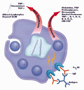
A mast cell is a normal part of the immune system and it a part of the immunological defense against invading organisms. These cells are attached to tissues that have an interface with the external world such as skin and they do not circulate around the body and respond to invaders. The mast cell possesses granules of special inflammatory biochemicals meant for use against invading parasites. This then is involved in a system with the IgE antibody which have the goal to sensitize the mast cell. This system works very well for defending against parasites; however, the IgE/mast cell system can be stimulated with other antigens that are of similar shape or size as parasitic antigens such as pollen which results in an allergic reaction.
A mast cell tumor is a tumor composed of numerous mast cells and it is a type of cancer despite its common name. This is due to the fact that a cancer is classified as a disease in which abnormal cells divide uncontrollably and destroy body tissue. Mast cell tumors are the most common form of skin tumors in dogs and make up more than 21% of the skin tumors. They are highly unstable and can degranulate or release their toxic granules with even a simple stimulus like a brief brush against it. These tumors can be relatively hard to treat due to their nature since a specific margin is unclear due to their degranulating nature. What is important is understanding the grades of these tumors since it influences the prognosis for your canine companion:
Stage 0: one tumor but incompletely excised from the skin. Stage I: one tumor confined to the skin with no regional lymph node involvement. Stage II: one tumor confined to the skin, but with regional lymph node involvement present. Stage III: many tumors or large, deeply infiltrating tumors, with or without lymph node involvement. Stage IV: any tumor with distant spread evident. In order to determine the tumor stage some probing of other lymphoid organs must be performed.
Low grade tumors with removal generally have a good prognosis and there tends to be a median life expectancy of your pet. However, once lymph node involvement has begun radiation or chemotherapy therapy is an option; however, life expectancy is greatly decreased due to the malignant nature of the cells. Also, due to the nature of the mast cells and histamine being constantly released all pets with this diagnosis, be it low grade or high grade, are recommended to be put on an antihistamine to help reduce inflammation.

The diagnosis of a mast cell tumor can be rather confusing and overwhelming, but armed with a little bit of knowledge and the reality of the diagnosis can make understanding it a bit easier if this issue ever arises for your canine companion. This issue can also afflict our feline companions, but it is a more common ailment for our canine companions!
